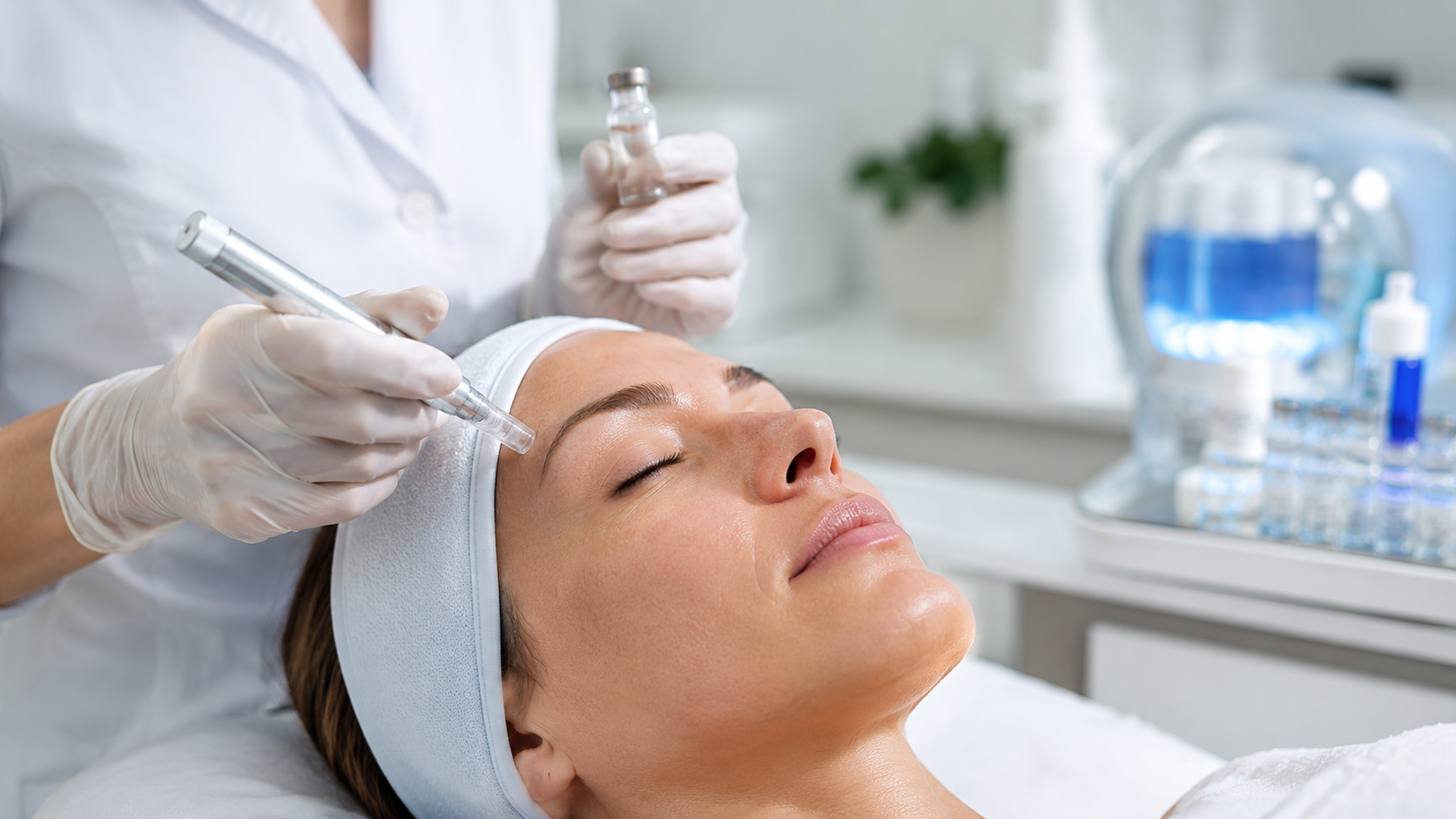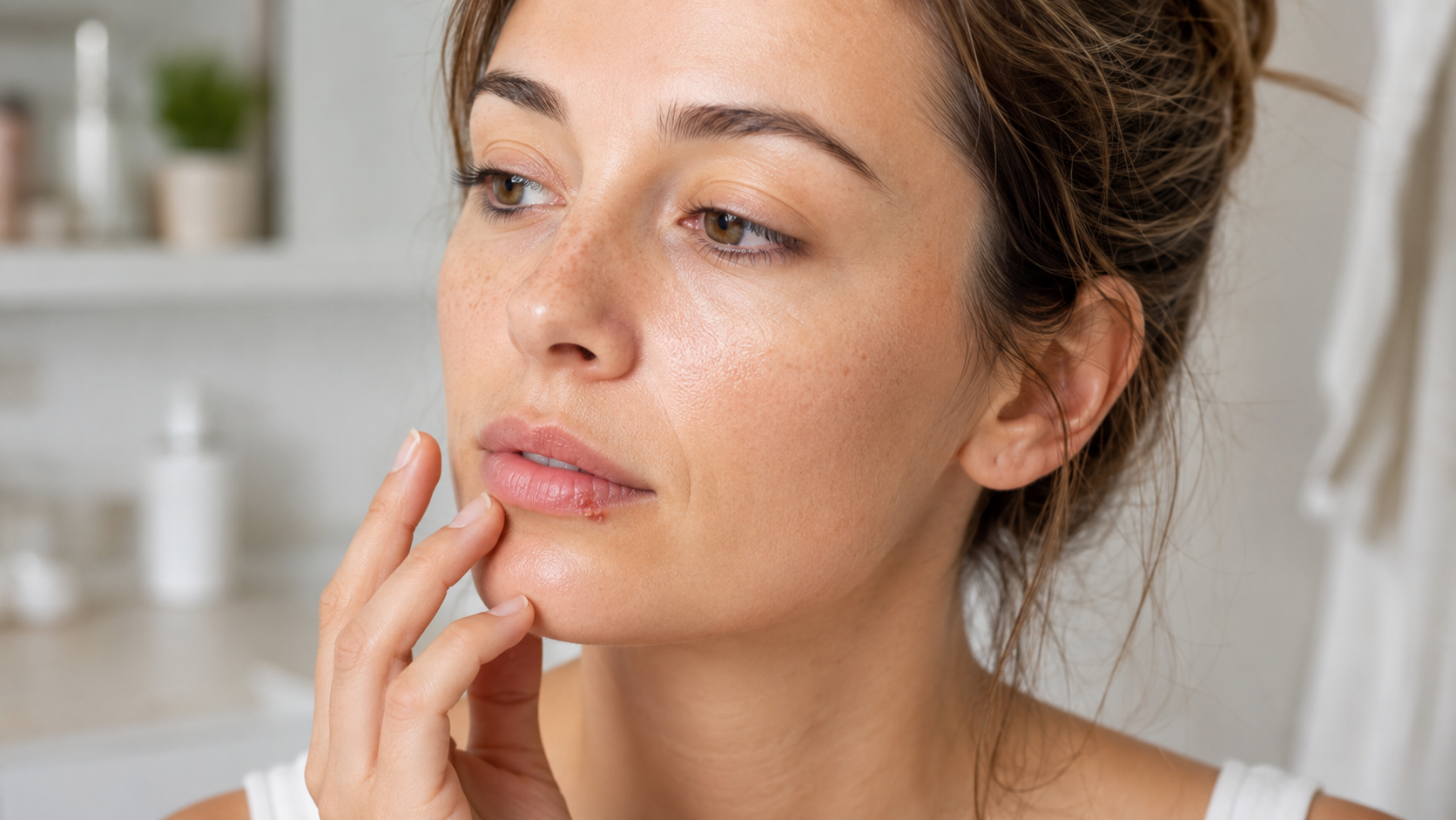

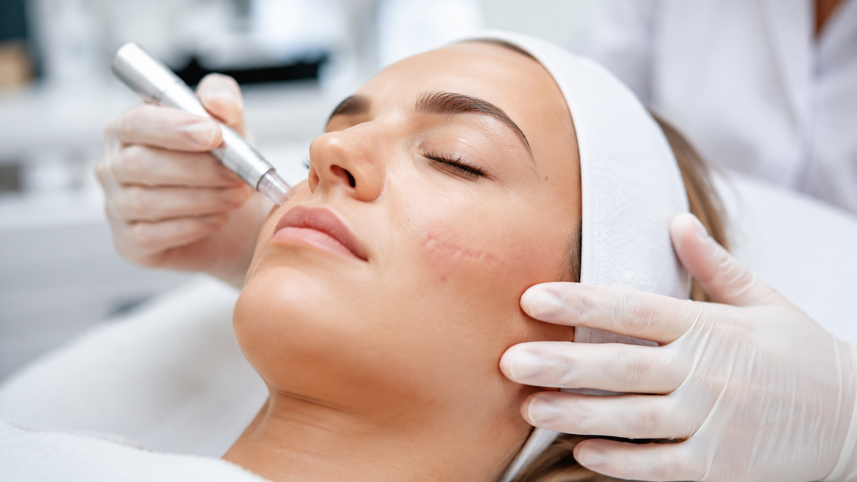
Scars form as a result of deeper skin injuries during the wound healing process. Depending on the depth of the injury, individual factors (skin type, predisposition, location) and aftercare, they may be flat and inconspicuous or raised, reddened and aesthetically troubling.
In the non-medical field, beauticians play an important role in accompanying scar care and supporting skin regeneration. One principle is clear here: cosmetics accompany – medicine treats.
Cosmetic treatments can support scar maturation, but only on scars that are completely healed, pain-free and non-irritated. Caring for fresh, inflamed or painful wounds, as well as invasive procedures (injections, lasers, medical needling), is the responsibility of a physician.
Scar formation: scars only form when the skin is injured at least as far as the dermis and the tissue is replaced by scarred connective tissue (see box). Superficial injuries to the epidermis heal without scarring.
Box: brief outline of wound healing in scar formation
- Exudation phase: blood and tissue fluid cleanse the wound, coagulation stops the bleeding, and fibrin (the “wound-closing protein”) seals it provisionally.
- Cleansing or resorption phase: immune cells remove tissue debris and pathogens and initiate repair.
- Proliferation phase: connective tissue cells form collagen, new vessels grow in, and granulation tissue develops.
- Repair or epithelialisation phase: the tissue consolidates, the wound surface closes – a scar is formed.
Scar tissue differs markedly from healthy skin: the collagen fibres run in parallel and elastic fibres are largely absent. The scar is less elastic and has no hairs, pigment cells, sebaceous glands or sweat glands. Remodelling processes continue even after wound healing is complete: collagen fibres become denser, the scar may shrink or harden, and blood flow decreases. Initially reddish scars fade over time.
A distinction is made between atrophic scars, hypertrophic scars and keloids, with treatment always guided by the type of scar. The degree of maturity (mature vs. immature) also plays a role.
Mature scars are pale, level with or minimally above the surrounding skin, soft and painless. Immature scars are still slightly raised, reddened and may itch. Depending on individual predisposition, location, cause and healing progression, however, more conspicuous scars can also develop.
- Atrophic scars lie below the level of the skin and result from tissue loss or insufficient collagen formation during wound healing. Typical examples are acne or chickenpox scars. A distinction is made between narrow, deep “ice-pick” scars, punched-out “boxcar” scars and broader “rolling” scars. They occur above all after inflammatory, sometimes deep acne and appear mainly on the face and back.
- Hypertrophic scars are raised, sometimes ridged, reddened and confined to the area of injury. They result from an excessive healing process with increased connective tissue formation, encouraged by tensile stress, delayed wound healing or inflammation. Itching, pain and functional limitations – particularly over joints – are possible. Spontaneous regression is possible but rare.
- Keloids grow beyond the boundaries of the original wound. They are firm, ridged, reddish to brownish, and often painful or itchy. They occur preferentially in people with a genetic predisposition and in darker skin types, frequently on the upper half of the body (presternal area, shoulder region/upper back, upper arms, earlobes), and always require medical treatment. Unlike hypertrophic scars, they grow progressively.
Complete removal of scars is not possible with cosmetic methods. The aim is a visible and palpable improvement by promoting regeneration, increasing elasticity, reducing hardening and feelings of tightness, and visually refining the appearance of the skin. The sooner treatment begins after wound closure, the better the prospects of success. Keloids must always be left to dermatologists.
Cosmetic options include manual techniques such as scar massage or lymphatic techniques. These support blood circulation and metabolism, improve the elasticity of the scar tissue and reduce congestion and feelings of tightness. Where devices are used, the rule is: non-invasive only, and only after complete wound closure.
Ultrasound works with high-frequency sound waves that generate micro-vibrations and thermal effects in the tissue. The circulation-boosting effect and the stimulation of metabolism can help to loosen the tissue and the scar and make them more supple. The absorption of regeneration-promoting active ingredients can also be improved.
Peeling treatments – herbal peeling courses, for example – can support the regeneration of scar tissue and make the scar relief appear smoother, so that the scar looks flatter, softer and less visible. Not all exfoliation methods (chemical peels, for instance) are equally suitable for scar tissue, however.
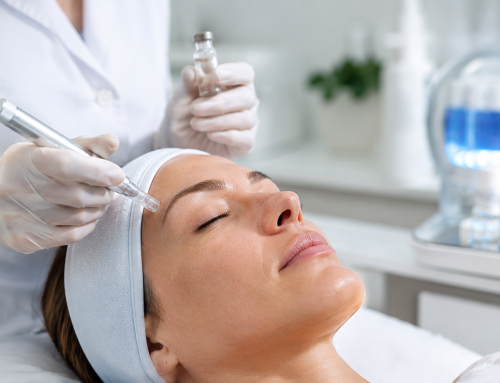
Other superficial procedures, such as microdermabrasion (mechanical skin resurfacing), can visually improve superficial atrophic scars. Depending on the initial condition, repeated treatments are required.
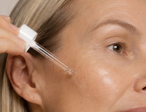
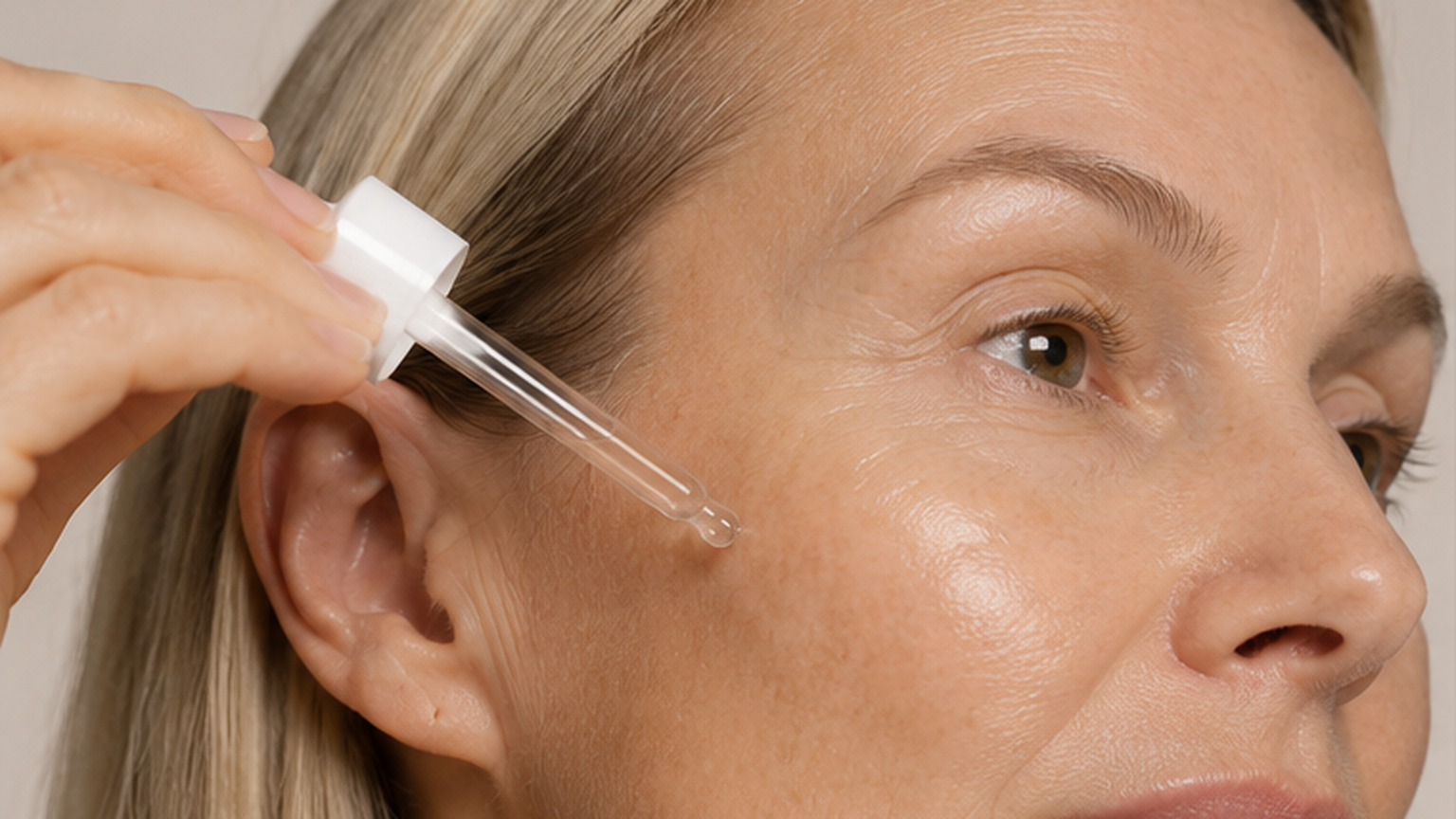
Cosmetic scar care and active ingredients: up until wound closure – as long as no permanent scar has yet formed and moist granulation tissue is still present – panthenol supports the wound healing process and the moisture balance. Ceramides strengthen the skin barrier, while allantoin, niacinamide, ectoin and madecassosides (Centella asiatica) can stimulate the skin’s own regeneration. Natural oils such as squalane keep the scar tissue supple and protect it from drying out; urea can relieve itching.
Since scars have no pigment cells, consistent light protection (SPF 50) is important for at least the first three months. Sunburn and subsequent complications (hyperpigmentation, development of skin cancer) must be avoided.
Silicone preparations – as a gel, cream or sheet – are of great importance. Applied early, regularly and for at least three months, they can visibly improve the appearance of a scar. The sealing (occlusive) film increases the hydration of the scar and thus supports scar maturation. Sheets should ideally be worn continuously (except when showering). Gel or cream is applied in a thin layer. Tip: do not massage it in, as this causes the silicone film over the scar to break up and the effect is lost.
As an alternative – over joints, for example, where the silicone film is more easily broken up by movement – preparations containing allantoin, heparin (sodium) and onion extract are better suited for scar care.
Among medical treatment methods, surgical corrections can improve scars but not remove them completely. Surgery should only be performed once scar maturation is complete (usually after at least 12 months). Excessive scarring calls for a clear distinction: hypertrophic scars are often readily treatable, whereas keloids are assessed far more cautiously because of their high risk of recurrence, particularly after surgical intervention. Treatment is therefore planned individually by a physician – usually starting with gentle procedures and moving on to more intensive methods if required.
Cryotherapy refers to freezing with liquid nitrogen and is frequently used for hypertrophic scars. After freezing, pigment disorders may occur due to the destruction of cold-sensitive melanocytes, along with slight blistering. Repeat sessions at intervals of around four weeks are generally required to achieve the desired effect.
Corticosteroids (cortisone) are injected directly into the scar. This inhibits collagen formation and flattens the scar. Sessions should be repeated every four weeks. This works particularly well on hypertrophic scars and active, bright red keloids. In the latter case, the injection is often combined with cryotherapy, since the tissue is temporarily oedematous after freezing, which makes application easier.
Laser procedures are becoming increasingly important in scar therapy and are used selectively depending on the type of scar. Laser treatment of scar tissue belongs in the hands of experts, since an overly aggressive approach can even make scars worse.
Vascular lasers such as pulsed dye lasers (PDL) are used on reddened, excessive scars and in some cases preventively on still-fresh scars. By obliterating the small blood vessels, the redness of the scar decreases. In addition, the tissue becomes undersupplied, which can lead to flattening of the scar. Efficacy is limited in darker skin types; overall, moderate to good results are usually achieved.
Ablative lasers such as CO2 or Er:YAG lasers are used on hypertrophic and mature scars as well as on acne scars. The aim is to level the surface of the scar and bring it back into line with the surrounding skin. Thanks to its thermal effect, the CO2 laser can additionally support tissue remodelling, which can visibly improve scar quality.
Further combination procedures include laser treatment followed by autologous blood therapy (PRP, platelet-rich plasma), which optimises the healing process and treatment results. Another is laser-assisted drug delivery (LADD), in which the action of lasers is combined with the subsequent application of medical or cosmetic active ingredients: the laser creates tiny channels in the skin through which these substances can then penetrate better and more deeply.